2D/3D spheroid culture
Topographical substrates (product type G0)
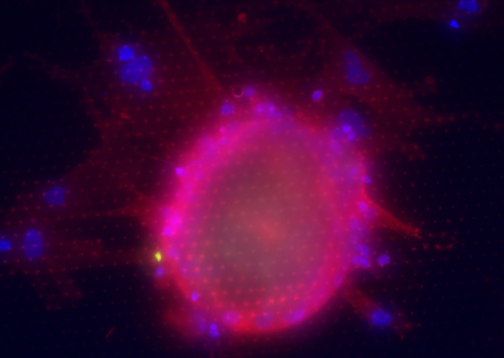
In order to form spheroids from cells that grow naturally in cell clusters, it is sufficient to seed 10,000-20,000 cells on the UV sterilized 1x1 cm template in a 6- (for three templates) or 24-well (for a single template) plate. After an incubation time, allowing the cells to attach, the well is filled with a cell culture medium. The cells start to settle from the first day in clusters and will grow to dense spheroids of approximately 100-150 µm in 1-6 days, depending on the cell line. Cells that naturally migrate, such as invading cancer-associated fibroblast, will grow in 2D. No further treatment or detachment is necessary. Cells can be fixed, stained, and microscopically visualized on the Encytos template.
Complex Tumor Spheroid Formation and One-Step Cancer-Associated Fibroblasts Purification from Hepatocellular Carcinoma Tissue Promoted by Inorganic Surface Topography
Abstract: In vitro cell models play important roles as testbeds for toxicity studies, drug development, or as replacements in animal experiments. In particular, complex tumor models such as hepatocellular carcinoma (HCC) are needed to predict drug efficacy and facilitate translation into clinical practice.
In this work, topographical features of amorphous silicon dioxide (SiO2) are fabricated and tested for cell culture of primary HCC cells and cell lines. The topographies vary from pyramids to octahedrons to structures named fractals, with increased hierarchy and organized in periodic arrays (square or Hexagonal). The pyramids were found to promote complex 2D/3D tissue formation from primary HCC cells. It was found that the 2D layer was mainly composed of cancer-associated fibroblasts (CAFs), while the 3D spheroids were composed of tumor cells enwrapped by a CAF layer. Compared with conventional protocols for 3D cultures, this novel approach mimics the 2D/3D complexity of the original tumor by invading CAFs and a microtumor. Topographies such as octahedrons and fractals exclude tumor cells and allow one-step isolation of CAFs even directly from tumor tissue of patients as the CAFs migrate into the structured substrate. Cell lines form spheroids within a short time. The presented inorganic topographical surfaces stimulate complex spheroid formation while avoiding additional biological scaffolds and allowing direct visualization on the substrate.