Fast cell differentiation
Topographical substrates (Product type G0-G4)
Cell differentiation from stem cells or other (de-differentiated) cells able to differentiate is often time-consuming and/or requires growth factors. The Encytos templates induce differentiation without factors into different lineages.
1) Adipose-derived stem cells show after 1-day nestin-positivity indicating neurosphere features on G1-G4.
2) HT29 differentiates in 6 days without methotrexate in standard medium mainly into Paneth cells on G0 and G1. Moreover, we observed much faster within 2 days mucus release.
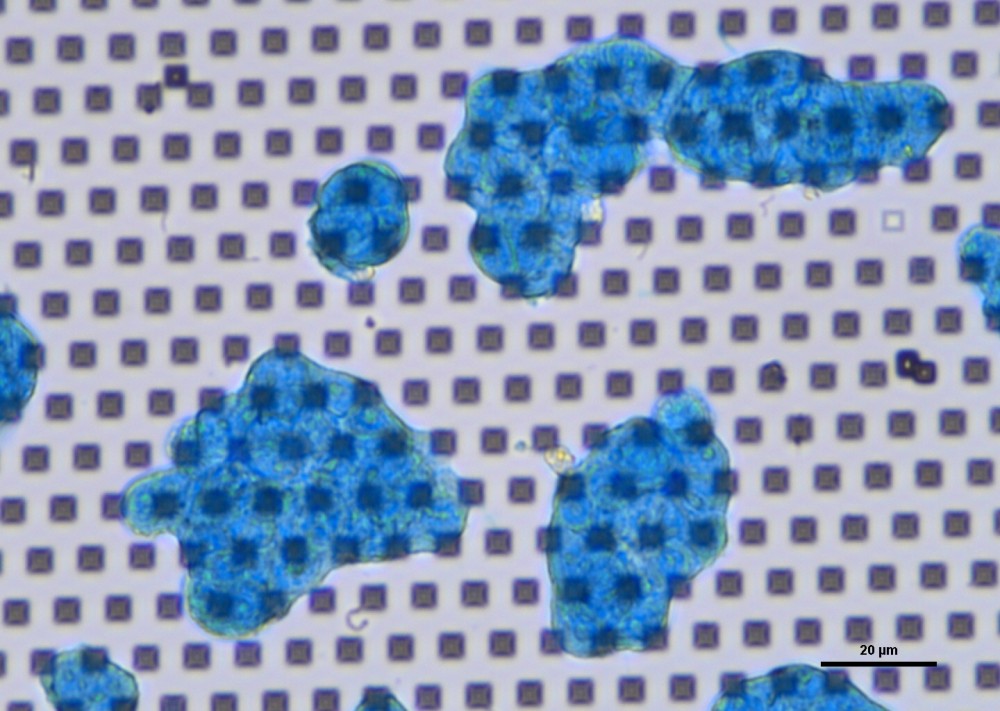
Figure: Mucus-production of HT29 cells after 4 days on G1.
The Fast Track for Intestinal Tumor Cell Differentiation and In Vitro Intestinal Models by Inorganic Topographic Surfaces
Abstract: Three-dimensional (3D) complex in vitro cell systems are well suited to providing
meaningful and translatable results in drug screening, toxicity measurements, and biological
studies. Reliable complex gastrointestinal in vitro models as a testbed for oral drug administration
and toxicity are very valuable in achieving predictive results for clinical trials and reducing animal
testing. However, producing these models is time-consuming due to the lengthy differentiation of
HT29 or other cells into mucus-producing goblet cells or other intestinal cell lineages. In the present
work, HT29 cells were grown on an inorganic topographic surface decorated with a periodic pattern
of micrometre-sized amorphous SiO2 structures for up to 35 days. HT29 cells on topographic
surfaces were compared to undifferentiated HT29 in glucose-containing medium on glass or culture
dish and with HT29 cells differentiated for 30 days in the presence of methotrexate
(HT29-MTX). The cells were stained with Alcian blue for mucus, antibodies for mucus 2 (goblet
cells), villin (enterocytes), lysozyme (Paneth cells), and FITC-labeled lectins to identify different
cells, glycomic profiles, and cell features. We observed that HT29 cells on topographic surfaces
showed more similarities with the differentiated HT29-MTX than with undifferentiated HT29.
They formed islands of cell clusters, as observed for HT29-MTX. Already after 2 days, the first
mucus secretion was shown by Alcian blue stain and FITC-wheat germ agglutinin. After 4–6 days,
mucus was observed on the cell surface and in the intercellular space. The cell layer was undulated,
and in 3D reconstruction, the cells showed a clear polarisation with a strong actin signal to one
membrane. The lectins and the antibody-staining confirmed the heterogeneous composition of
differentiated HT29 cells on topographic surfaces after 6–8 days, or after 6–8 days following MTX
differentiation (30 days).